In July 2026, the research group led by Ouyang Liangliang at Tsinghua University published a seminal biofabrication study in Nature, introducing a new 3D printing method based on sub‑voxel oriented fiber architecture.
Through an in‑situ fiber transformation strategy, the technique generates highly aligned, continuously uniform sub‑voxel hydrogel fiber bundles (5–30 µm in diameter) directly inside conventionally extruded hydrogel filaments. These cell‑scale fibers effectively guide the oriented growth of skeletal muscle cells, enabling the formation of the longest myotube structures ever reported in vitro—averaging 1.1 mm and reaching up to 3 mm—while also demonstrating strong potential for in‑vivo tissue repair.
Two Long‑Standing Industry Pain Points
Tissues such as skeletal muscle, peripheral nerves, and blood vessels all rely on highly ordered anisotropic fiber architectures to perform their physiological functions. However, conventional extrusion‑based bioprinting has long been constrained by two critical shortcomings. First, it is difficult to generate uniform microfibers that match the size of mammalian cells (5–30 µm) inside printed strands; pre‑mixing short fibers results in random arrangements, limited length, and poor controllability. Second, there is an inherent trade‑off between ink fluidity and shape fidelity after printing. Alternatives like coaxial nozzles or electrospinning either increase equipment costs, narrow the range of compatible materials, or fail to directly print 3D constructs with living cells.
SHIFT Breaks the Deadlock: Temperature‑Controlled Stretching Turns Droplets into Fibers
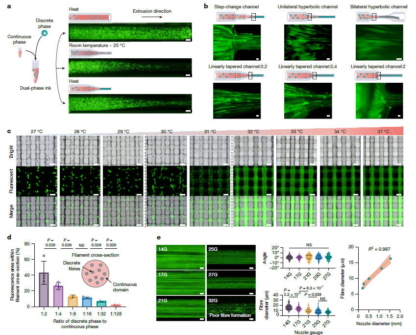
The team designed a "discrete‑continuous" biphasic ink system that exploits the opposite thermosensitive behavior of its two components—upon heating, the pectin‑based discrete phase transitions from gel to sol, while the gelatin‑based continuous phase turns into a micellar state, providing physical confinement. During extrusion through a contracting channel, the elongational flow field stretches the liquid droplets over 10,000‑fold along the extrusion direction, transforming them from spheres into continuous microfibers, which are then instantly locked in place by the rapid self‑recovery of the matrix. This process is largely insensitive to nozzle geometry and operates stably above 32 °C within a 27–37 °C window, making it compatible with standard commercial bioprinters.
Flexible Control and Broad Compatibility
The nozzle inner diameter shows a strong linear correlation with fiber diameter (R² = 0.987), and extrusion rates from 0.1 to 10 mL/min have negligible impact on fiber orientation. The ink system can be flexibly formulated with particles, microspheres, or even pre‑crosslinked gels of different compositions, allowing multi‑component sub‑voxel fibers to be arranged in parallel through a single nozzle. Gelatin fibers can also be dissolved out to create interconnected microchannels. The technique works with a wide range of hydrogels—including alginate, methacrylated hyaluronic acid, PEGDA, and silk fibroin—and can produce conductive composite fibers, with the elastic modulus of the printed constructs increased by 2–3 times over conventional hydrogels.
Three Application Scenarios and In‑Vivo Validation
The technology also enables a standalone spinning mode for high‑throughput collection of pure hydrogel microfibers. Three major applications have achieved key breakthroughs: sacrificial gelatin fibers form capillary‑scale networks after dissolution; microfiber‑embedded iPSC spheroids create internal transport pathways that markedly reduce cell death at the core of organoids; and myoblast‑laden scaffolds cultured in vitro produce ultra‑long myotubes that exhibit spontaneous rhythmic contractions for up to 60 days, with an amplitude of 267 µm.
In animal models, an 8‑week implantation of the cell‑laden SHIFT scaffolds into mouse volumetric muscle loss defects led to newly formed myofibers approaching healthy control levels, along with substantial recovery of grip strength and endurance. In a rat sciatic nerve defect model with a 10‑mm gap, the SHIFT fiber scaffolds guided oriented axonal regrowth across the lesion, achieving regeneration results comparable to autologous nerve grafting.
Future Outlook
The SHIFT process requires no sophisticated or custom‑built hardware and can be directly integrated into existing 3D printing and microfiber spinning systems. Its core strategy—coupling elongational flow with inverse phase transition for in‑situ fiber formation—is theoretically extensible to other stimuli‑responsive systems, such as photo‑ or acoustic‑sensitive materials. The hydrogel microfibers produced by SHIFT can serve as versatile building blocks for fiber‑reinforced printing inks, sacrificial vascular templates, and internal molecular transport channels in organoids, opening up broad possibilities for the processing of anisotropic soft materials and biofabrication.